



Somewhere between the walking sticks and the ophthalmologist’s verdict, Rhys Hibbert, 48, from Bedfordshire, ran out of good options. A pituitary adenoma — roughly 11 mm, small enough to miss on a bad day, large enough to crush his optic nerves — was steadily erasing his peripheral vision. Without surgery, permanent blindness was the destination. In May 2026, surgeons at London’s National Hospital for Neurology and Neurosurgery removed the tumor using a real-time AI system analyzing live surgical video. It’s a world first, and it matters well beyond one man’s recovery.
What the AI Actually Does in the Operating Room
A camera threaded through the nose, an algorithm watching every frame — here’s how this system actually works inside the skull.
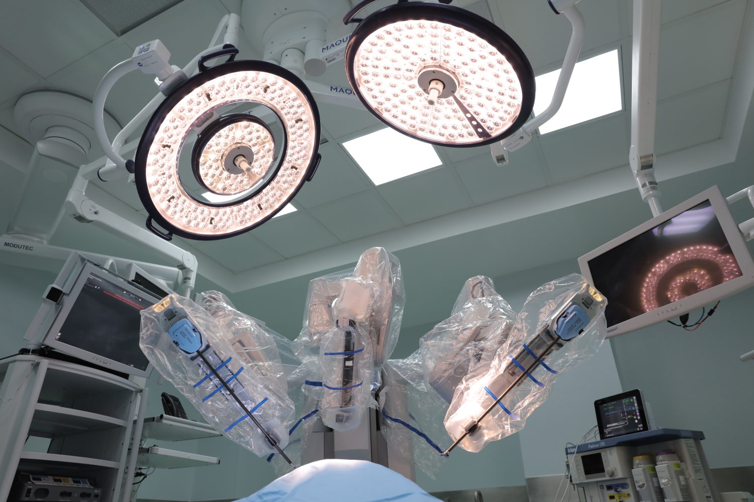
Your phone’s face-unlock recognizes your face by pattern. This system does something similar, except instead of faces, it recognizes anatomy that, if nicked, causes blindness or stroke. Developed at UCL’s Hawkes Institute under Dr. Sophia Bano, the system watches live endoscopic video and color-codes critical structures — optic nerves, carotid arteries, major vessels — on a secondary screen beside the surgeon. The scalpel stays in human hands. The AI makes sure those hands know exactly what’s nearby.
A few facts worth anchoring:
- Trained on hundreds of annotated pituitary surgery videos — more procedures than most surgeons accumulate in a career
- Runs on NVIDIA Clara IGX, a medical-grade edge AI platform processing video locally (hospital internet, the research notes, is unreliable)
- Part of a formal IDEAL Stage 1–2a clinical trial at NHNN; Hibbert’s case is among the first six feasibility cases
- Funded by NIHR and Google, with hardware from NVIDIA
“A second expert pair of eyes.” — Prof. Hani Marcus, consultant neurosurgeon, UCL
What Happened to Rhys Hibbert
From walking aids and collapsing vision to independence in under a week — the outcome is as striking as the technology behind it.
Before the operation: walking aids, progressive vision loss, hormone disruption at 48. After: within a week, Hibbert walked independently without glasses or sticks. At eight weeks, he described seeing “in 360 degrees.” Prof. Marcus and resident Danyal Khan made every cut. The AI flagged danger zones on a secondary screen. That distinction matters.
“Designed to help recognise critical anatomy, surgical instruments and tissue interactions in real time, supporting the surgeon during highly delicate procedures.” — Dr. Sophia Bano, UCL Hawkes Institute
“Assistive, not autonomous” is the honest framing. The surgeon retains full control; the AI offers cues, not commands. This is still early-stage technology — six feasibility cases, broad regulatory approval pending. The system improves anatomy recognition, according to peer-reviewed research from the same UCL group, but it doesn’t eliminate the need for surgical expertise and vigilance. A mislabeled structure, in a space this confined, isn’t an abstract risk.
Future versions are expected to track instruments in real time and overlay pre-operative imaging mid-procedure — closer to a true co-pilot than a color-coded map. An algorithm trained in months now sees an operating field with more accumulated experience than most human surgeons ever will. That’s worth sitting with.