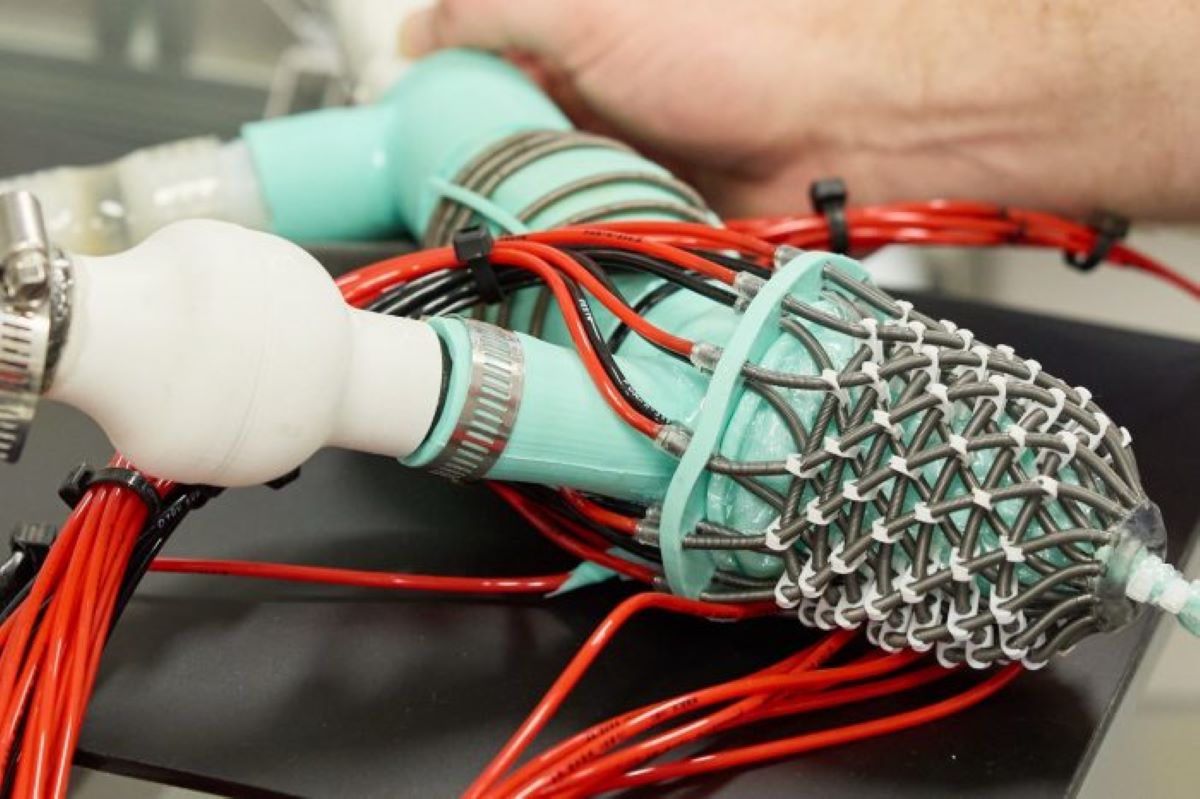
Every cardiac device headed into a patient’s chest was tested somewhere first. Until now, that “somewhere” was often a rigid plastic chamber with a simplified valve — or a living animal. Neither tells the full story. Researchers at UNSW Sydney have built something more honest: a fully synthetic soft robotic left heart that reproduces the mitral valve, papillary muscles, and the tiny tendon-like chordae tendineae, all in silicone and hydraulic muscle. This isn’t going inside anyone. It’s a bench-top simulator designed to stress-test the tools that do.
Built to Break
Layered hydraulic muscles replicate the twist and squeeze of a real ventricle, giving device makers a testbed that actually moves like human tissue.
Silicone membranes form compliant inner chambers while hydraulic artificial muscles wrap around them, arranged in the same layered fibre geometry as ventricular myocardium. The result, published in Advanced Science, is a model that twists and shortens during contraction the way a human heart does — not the way a piston approximates it. Think Formula 1 teams simulating race conditions in a wind tunnel before the car touches a track. Same logic, dramatically higher stakes.
The simulator’s programmable capabilities include:
- Adjustable papillary muscle tension to simulate mitral valve prolapse and regurgitation
- Hemodynamic waveforms matching a healthy human left heart, confirmed with invasive sensors
- Compatibility with clinical echocardiography — regurgitant jets visible on ultrasound
- Programmable compliance to replicate HFpEF, a form of heart failure affecting roughly half of all cases
- Geometry customizable from patient imaging data, including echocardiography and CT
When researchers increase papillary muscle tension, leaflets fail to close properly and blood flows backward — mitral regurgitation, reproduced at a bench. “Build realistic artificial heart models that can help researchers understand disease and develop safer, more effective devices before they are tested on animals or reach patients,” says A/Prof Thanh Nho Do, who leads the UNSW team.
The Stakes Beyond the Lab
From modeling a poorly understood form of heart failure to reducing early-stage animal testing, the simulator’s ambitions stretch well past the bench.
HFpEF — heart failure where the ventricle stiffens but still pumps — currently lacks targeted mechanical therapies. The simulator reproduces its hallmark diastolic dysfunction by programmatically reducing ventricular compliance, producing slower filling and elevated pressures that mirror early patient data, according to the team’s Nature Communications paper. Do argues HFpEF “deserves its own mechanical treatment options,” and a platform that can reliably replicate it may be what finally gets that conversation moving.
A soft robotic catheter was already navigated inside the beating model, detecting contact with valve leaflets in real time. Scientia Professor Nigel Lovell noted that “ultrasound imaging from the model resembled human cardiac imaging owing to the biomimetic form and function.” Device companies could eventually test valve clips, annuloplasty rings, and catheters under realistic hemodynamic conditions here — before any animal sees them.
The honest caveat: this remains proof-of-concept. Extensive validation against diverse patient datasets is still required. But the trajectory points somewhere specific — build a physical model from a patient’s CT scan, stress-test three valve sizes before the surgeon picks up a scalpel. That’s where this is heading.